Cell Meter Beta-Arrestin蛋白质易位GPCR信号检测试剂盒 货号36391-AAT Bioquest荧光染料

上海金畔生物科技有限公司代理AAT Bioquest荧光染料全线产品,欢迎访问AAT Bioquest荧光染料官网了解更多信息。

Cell Meter Beta-Arrestin蛋白质易位GPCR信号检测试剂盒
Cell Meter Beta-Arrestin蛋白质易位GPCR信号检测试剂盒

Cell Meter Beta-Arrestin蛋白质易位GPCR信号检测试剂盒    货号36391 货号 36391 存储条件 在零下15度以下保存, 避免光照
规格 200 Tests 价格 11628
Ex (nm) Em (nm)
分子量 溶剂
产品详细介绍

简要概述

所有G蛋白偶联受体(GPCR)在通过配体结合激活后,都会通过一条共同途径迅速发生脱敏作用。 β-arrestin(一种细胞质蛋白)与激活的受体的结合使GPCR信号失活,并启动了受体向细胞内的易位,在此细胞中配体被去除,受体被循环回到细胞膜。通过将荧光标记(例如GFP)附着到β-arrestin,可以检测受体arrestin复合物的位置。由于脱敏仅发生在激活的受体上,因此检测β-arrestin的转运和随后的受体再循环提供了检测GPCR靶标激活的可靠方法。 Cell Meter Beta-Arrestin蛋白易位GPCR信号试剂盒提供了功能强大的功能分析,可通过荧光成像筛选目标化合物针对已知或孤立GPCR靶标的活性。靶向GPCR的激活诱导荧光向细胞膜和内吞囊泡的移位。


适用仪器


荧光显微镜  
激发: FITC滤波片
发射: FITC滤波片
推荐孔板: 黑色透明底板

产品说明书

样品实验方案

概述

准备细胞进行转染
准备Transfectamine 5000-DNA混合物
将Transfectamine 5000-DNA混合物添加到细胞培养物中,并孵育过夜
转染后24-30小时将转染的细胞转移到96孔板中,并将培养物孵育过夜
在荧光显微镜下分析由GPCR激活引起的转运


细胞准备

细胞密度在转染时它们将达到约60-70%的融合度。
转染前用新鲜的生长培养基替换。
注意:例如,对于6孔板,每孔替换为2 mL培养基,对于10 cm板,则替换为6 mL培养基。


储备溶液配制

除非另有说明,否则所有未使用的储备溶液应分为一次性使用的等分试样,并在制备后储存在-20°C下。避免重复冻融循环。

β-arrestin-GFPDNA储备溶液
向小瓶β-arrestin-GFPDNA(组分A)中加入10 µL ddH2O,充分混合至终浓度为1 µg / µL。


操作步骤

1.将3 µg DNA(例如1.5 µg Beta-arrestin-GFP DNA储备液和1.5 µg您准备的GPCR DNA)与200 µL无血清培养基混合。
2.向混合物中加入9 µL Transfectamine 5000(组分B)。
3.充分混合并在室温下孵育20分钟。
注意:Transfectamine 5000和DNA的比例需要针对不同的细胞系进行优化,通常,在我们的测试中,Transfectamine 5000转染试剂(µL)与DNA(µg)的比例应为3-5 µL:1ug。

表1. 6孔板和10 cm板的样本表

  6孔板(每孔) 10cm板
新鲜培养基 2 mL 6 mL
质粒 3 µg 10 µg
无血清培养基 200 µL 600 µL
Transfectamine 5000转染试剂 ~9 µL ~30 µL


转染和转运方案
1.将Transfectamine 5000 -DNA混合物添加到培养板中,并孵育过夜。
注意事项:最早在转染后16小时即可检测到重组蛋白。转染后72〜96小时可观察到最大表达水平。
2.转染后24-30小时将转染的细胞转移到96孔板中,并孵育过夜。
使用FITC滤光片(Ex / Em = 488/530 nm)在荧光显微镜下检测受体激活诱导的β-arrestin易位。


图示
Cell Meter Beta-Arrestin蛋白质易位GPCR信号检测试剂盒    货号36391

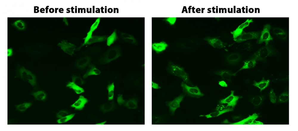
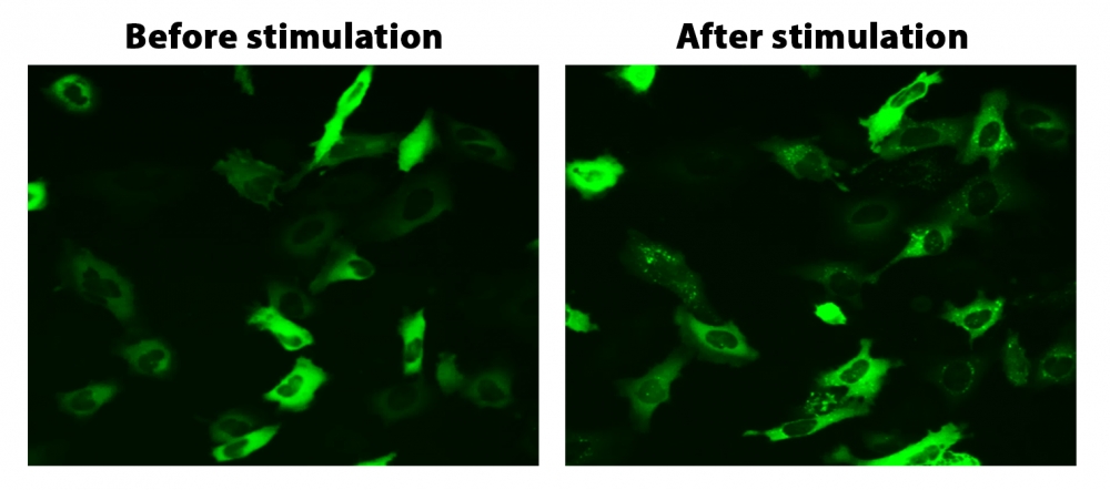
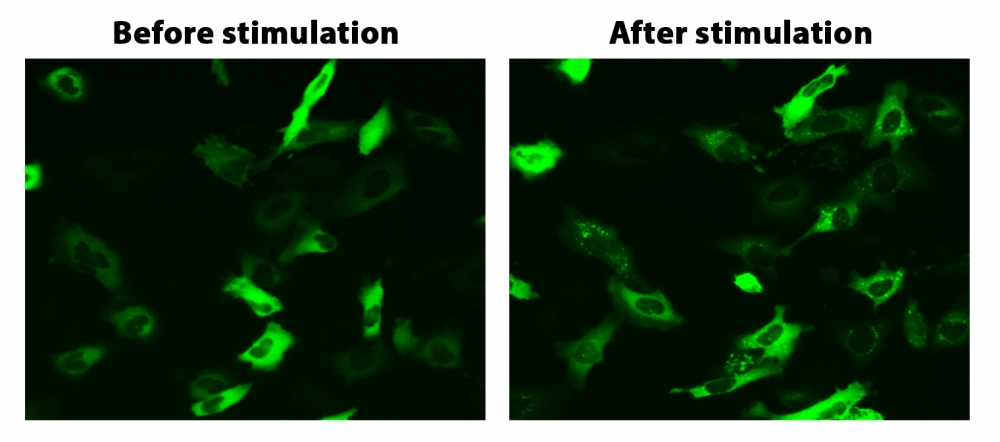
图1. HeLa细胞中β-arrestin的易位。 HeLa细胞用β-arrestin-GFP和加压素受体2(V2R)瞬时转染。 HeLa细胞在6孔板中培养,并融合至〜60%。用9 µL Transfectamine 5000转染等量的β-arrestin-GFP(1.5 µg)和V2R质粒(1.5 µg)。转染后约30小时,将细胞转移到96孔板中。转染后约48小时,将加压素(1 µM)加入细胞中以诱导β-arrestin-GFP易位。使用FITC通道在荧光显微镜下加压素处理之前和之后2小时拍摄图像。


参考文献

Screening cellular feature measurements for image-based assay development.
Authors: Logan, David J and Carpenter, Anne E
Journal: Journal of biomolecular screening (2010): 840-6

Kappa opioid receptor screen with the Tango beta-arrestin recruitment technology and characterization of hits with second-messenger assays.
Authors: Doucette, Christopher and Vedvik, Kevin and Koepnick, Elizabeth and Bergsma, Aaron and Thomson, Brian and Turek-Etienne, Tammy C
Journal: Journal of biomolecular screening (2009): 381-94

Multiplexed assays by high-content imaging for assessment of GPCR activity.
Authors: Ross, D A and Lee, S and Reiser, V and Xue, J and Alves, K and Vaidya, S and Kreamer, A and Mull, R and Hudak, E and Hare, T and Detmers, P A and Lingham, R and Ferrer, M and Strulovici, B and Santini, F
Journal: Journal of biomolecular screening (2008): 449-55

Comparison of G-protein coupled receptor desensitization-related beta-arrestin redistribution using confocal and non-confocal imaging.
Authors: Haasen, Dorothea and Wolff, Michael and Valler, Martin J and Heilker, Ralf
Journal: Combinatorial chemistry & high throughput screening (2006): 37-47

High-content screening of known G protein-coupled receptors by arrestin translocation.
Authors: Hudson, Christine C and Oakley, Robert H and Sjaastad, Michael D and Loomis, Carson R
Journal: Methods in enzymology (2006): 63-78

High-throughput confocal microscopy for beta-arrestin-green fluorescent protein translocation G protein-coupled receptor assays using the Evotec Opera.
Authors: Garippa, Ralph J and Hoffman, Ann F and Gradl, Gabriele and Kirsch, Achim
Journal: Methods in enzymology (2006): 99-120

The ligand-independent translocation assay: an enabling technology for screening orphan G protein-coupled receptors by arrestin recruitment.
Authors: Oakley, Robert H and Hudson, Christine C and Sjaastad, Michael D and Loomis, Carson R
Journal: Methods in enzymology (2006): 50-63

Development and validation of algorithms for measuring G-protein coupled receptor activation in cells using the LSC-based imaging cytometer platform.
Authors: Ozawa, Kazuo and Hudson, Christine C and Wille, Kirsten R and Karaki, Sachiko and Oakley, Robert H
Journal: Cytometry. Part A : the journal of the International Society for Analytical Cytology (2005): 69-76

Quantitative cell-based high-content screening for vasopressin receptor agonists using transfluor technology.
Authors: Ghosh, Richik N and DeBiasio, Richard and Hudson, Christine C and Ramer, Everett R and Cowan, Conrad L and Oakley, Robert H
Journal: Journal of biomolecular screening (2005): 476-84

The cellular distribution of fluorescently labeled arrestins provides a robust, sensitive, and universal assay for screening G protein-coupled receptors.
Authors: Oakley, Robert H and Hudson, Christine C and Cruickshank, Rachael D and Meyers, Diane M and Payne, Richard E and Rhem, Shay M and Loomis, Carson R
Journal: Assay and drug development technologies (2002): 21-30

说明书
Cell Meter Beta-Arrestin蛋白质易位GPCR信号检测试剂盒.pdf

Cell Meter Beta-Arrestin蛋白质易位GPCR信号检测试剂盒 货号36390-AAT Bioquest荧光染料

上海金畔生物科技有限公司代理AAT Bioquest荧光染料全线产品,欢迎访问AAT Bioquest荧光染料官网了解更多信息。

Cell Meter Beta-Arrestin蛋白质易位GPCR信号检测试剂盒

Cell Meter Beta-Arrestin蛋白质易位GPCR信号检测试剂盒

Cell Meter Beta-Arrestin蛋白质易位GPCR信号检测试剂盒    货号36390 货号 36390 存储条件 在零下15度以下保存, 避免光照
规格 100 Tests 价格 9180
Ex (nm) Em (nm)
分子量 溶剂
产品详细介绍

简要概述

所有G蛋白偶联受体(GPCR)在通过配体结合激活后,都会通过一条共同途径迅速发生脱敏作用。 β-arrestin(一种细胞质蛋白)与激活的受体的结合使GPCR信号失活,并启动了受体向细胞内的易位,在此细胞中配体被去除,受体被循环回到细胞膜。通过将荧光标记(例如GFP)附着到β-arrestin,可以检测受体arrestin复合物的位置。由于脱敏仅发生在激活的受体上,因此检测β-arrestin的易位和随后的受体再循环提供了检测GPCR靶标激活的可靠方法。 Cell Meter Beta-Arrestin蛋白易位GPCR信号试剂盒提供了功能强大的功能分析,可通过荧光成像筛选目标化合物针对已知或孤立GPCR靶标的活性。靶向GPCR的激活诱导荧光向细胞膜和内吞囊泡的移位。


适用仪器


荧光显微镜  
激发: FITC滤波片
发射: FITC滤波片
推荐孔板: 黑色透明底板

产品说明书

样品实验方案

概述

准备细胞进行转染
准备Transfectamine 5000-DNA混合物
将Transfectamine 5000-DNA混合物添加到细胞培养物中,并孵育过夜
转染后24-30小时将转染的细胞转移到96孔板中,并将培养物孵育过夜
在荧光显微镜下分析由GPCR激活引起的转运


细胞准备

细胞密度在转染时它们将达到约60-70%的融合度。
转染前用新鲜的生长培养基替换。
注意:例如,对于6孔板,每孔替换为2 mL培养基,对于10 cm板,则替换为6 mL培养基。


储备溶液配制

除非另有说明,否则所有未使用的储备溶液应分为一次性使用的等分试样,并在制备后储存在-20°C下。避免重复冻融循环。

β-arrestin-GFPDNA储备溶液
向小瓶β-arrestin-GFPDNA(组分A)中加入10 µL ddH2O,充分混合至终浓度为1 µg / µL。


操作步骤

1.将3 µg DNA(例如1.5 µg Beta-arrestin-GFP DNA储备液和1.5 µg您准备的GPCR DNA)与200 µL无血清培养基混合。
2.向混合物中加入9 µL Transfectamine 5000(组分B)。
3.充分混合并在室温下孵育20分钟。
注意:Transfectamine 5000和DNA的比例需要针对不同的细胞系进行优化,通常,在我们的测试中,Transfectamine 5000转染试剂(µL)与DNA(µg)的比例应为3-5 µL:1ug。

表1. 6孔板和10 cm板的样本表

  6孔板(每孔) 10cm板
新鲜培养基 2 mL 6 mL
质粒 3 µg 10 µg
无血清培养基 200 µL 600 µL
Transfectamine 5000转染试剂 ~9 µL ~30 µL


转染和转运方案
1.将Transfectamine 5000 -DNA混合物添加到培养板中,并孵育过夜。
注意事项:最早在转染后16小时即可检测到重组蛋白。转染后72〜96小时可观察到最大表达水平。
2.转染后24-30小时将转染的细胞转移到96孔板中,并孵育过夜。
使用FITC滤光片(Ex / Em = 488/530 nm)在荧光显微镜下检测受体激活诱导的β-arrestin易位。


图示

Cell Meter Beta-Arrestin蛋白质易位GPCR信号检测试剂盒    货号36390

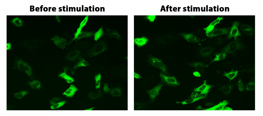
图1. HeLa细胞中β-arrestin的易位。 HeLa细胞用β-arrestin-GFP和加压素受体2(V2R)瞬时转染。 HeLa细胞在6孔板中培养,并融合至〜60%。用9 µL Transfectamine 5000转染等量的β-arrestin-GFP(1.5 µg)和V2R质粒(1.5 µg)。转染后约30小时,将细胞转移到96孔板中。转染后约48小时,将加压素(1 µM)加入细胞中以诱导β-arrestin-GFP易位。使用FITC通道在荧光显微镜下加压素处理之前和之后2小时拍摄图像。


参考文献

Screening cellular feature measurements for image-based assay development.
Authors: Logan, David J and Carpenter, Anne E
Journal: Journal of biomolecular screening (2010): 840-6

Kappa opioid receptor screen with the Tango beta-arrestin recruitment technology and characterization of hits with second-messenger assays.
Authors: Doucette, Christopher and Vedvik, Kevin and Koepnick, Elizabeth and Bergsma, Aaron and Thomson, Brian and Turek-Etienne, Tammy C
Journal: Journal of biomolecular screening (2009): 381-94

Multiplexed assays by high-content imaging for assessment of GPCR activity.
Authors: Ross, D A and Lee, S and Reiser, V and Xue, J and Alves, K and Vaidya, S and Kreamer, A and Mull, R and Hudak, E and Hare, T and Detmers, P A and Lingham, R and Ferrer, M and Strulovici, B and Santini, F
Journal: Journal of biomolecular screening (2008): 449-55

Comparison of G-protein coupled receptor desensitization-related beta-arrestin redistribution using confocal and non-confocal imaging.
Authors: Haasen, Dorothea and Wolff, Michael and Valler, Martin J and Heilker, Ralf
Journal: Combinatorial chemistry & high throughput screening (2006): 37-47

High-content screening of known G protein-coupled receptors by arrestin translocation.
Authors: Hudson, Christine C and Oakley, Robert H and Sjaastad, Michael D and Loomis, Carson R
Journal: Methods in enzymology (2006): 63-78

High-throughput confocal microscopy for beta-arrestin-green fluorescent protein translocation G protein-coupled receptor assays using the Evotec Opera.
Authors: Garippa, Ralph J and Hoffman, Ann F and Gradl, Gabriele and Kirsch, Achim
Journal: Methods in enzymology (2006): 99-120

The ligand-independent translocation assay: an enabling technology for screening orphan G protein-coupled receptors by arrestin recruitment.
Authors: Oakley, Robert H and Hudson, Christine C and Sjaastad, Michael D and Loomis, Carson R
Journal: Methods in enzymology (2006): 50-63

Development and validation of algorithms for measuring G-protein coupled receptor activation in cells using the LSC-based imaging cytometer platform.
Authors: Ozawa, Kazuo and Hudson, Christine C and Wille, Kirsten R and Karaki, Sachiko and Oakley, Robert H
Journal: Cytometry. Part A : the journal of the International Society for Analytical Cytology (2005): 69-76

Quantitative cell-based high-content screening for vasopressin receptor agonists using transfluor technology.
Authors: Ghosh, Richik N and DeBiasio, Richard and Hudson, Christine C and Ramer, Everett R and Cowan, Conrad L and Oakley, Robert H
Journal: Journal of biomolecular screening (2005): 476-84

The cellular distribution of fluorescently labeled arrestins provides a robust, sensitive, and universal assay for screening G protein-coupled receptors.
Authors: Oakley, Robert H and Hudson, Christine C and Cruickshank, Rachael D and Meyers, Diane M and Payne, Richard E and Rhem, Shay M and Loomis, Carson R
Journal: Assay and drug development technologies (2002): 21-30
说明书
Cell Meter Beta-Arrestin蛋白质易位GPCR信号检测试剂盒.pdf

荧光底物Beta-Ala-R110-ML 货号13205-AAT Bioquest荧光染料

上海金畔生物科技有限公司代理AAT Bioquest荧光染料全线产品,欢迎访问AAT Bioquest荧光染料官网了解更多信息。

荧光底物Beta-Ala-R110-ML

荧光底物Beta-Ala-R110-ML

荧光底物Beta-Ala-R110-ML    货号13205 货号 13205 存储条件 在零下15度以下保存, 避免光照
规格 1 mg 价格 1740
Ex (nm) 500 Em (nm) 522
分子量 628.56 溶剂 DMSO
产品详细介绍

简要概述

产品基本信息

货号:13205

产品名称:荧光底物Beta-Ala-R110-ML

规格:1mg

储存条件:-15℃避光防潮

保质期:12个月


产品物理化学光谱特性

分子量:628.56

外观:固体

溶剂:DMSO

激发波长(nm):500

发射波长(nm):522


产品介绍

Beta-Ala-R110-ML 是一种荧光底物,可用于检测氨肽酶 M 底物、丙氨酰氨肽酶和胰蛋白酶活性。 Beta-Ala-R110-ML 可用于通过检测其丙氨酰氨基肽酶活性来检测食品和其他样品中的某些细菌。与 Ala-AMC (#13458) 相比,Beta-Ala-R110-ML 更敏感。它的酶产物可以很容易地用普通的 FITC 通道或 FITC 滤波片组检测到。

点击查看光谱


参考文献

Design, synthesis and biological evaluation of substituted 3-amino-N-(thiazol-2-yl)pyrazine-2-carboxamides as inhibitors of mycobacterial methionine aminopeptidase 1.
AuthorsJuhás, Martin and Pallabothula, Vinod S K and Grabrijan, Katarina and Šimovičová, Martina and Janďourek, Ondřej and Konečná, Klára and Bárta, Pavel and Paterová, Pavla and Gobec, Stanislav and Sosič, Izidor and Zitko, Jan
JournalBioorganic chemistry (2022): 105489
A Potent Inhibitor of Aminopeptidase P2 Reduces Reperfusion Injury in Models of Myocardial Infarction and Stroke.
AuthorsLenz, Morgan Rae and Tsai, Shih-Yen and Roessler, Anne E and Wang, Yang and Sethupathi, Periannan and Jones, Walter Keith and Kartje, Gwendolyn L and Simmons, William Howard
JournalThe Journal of pharmacology and experimental therapeutics (2022)
The dual-targeted prolyl aminopeptidase PAP1 is involved in proline accumulation in response to stress and during pollen development.
AuthorsGhifari, Abi S and Teixeira, Pedro F and Kmiec, Beata and Singh, Neha and Glaser, Elzbieta and Murcha, Monika W
JournalJournal of experimental botany (2022): 78-93
Characterization and heterologous expression of a novel Co2+-dependent leucyl aminopeptidase Amp0279 originating from Lysinibacillus sphaericus.
AuthorsZhao, Puying and Zhang, Meng and Wan, Xiaofu and Geng, Peiling and Xiong, Hairong and Hu, Xiaomin
JournalApplied microbiology and biotechnology (2022)
KBE009: A Bestatin-Like Inhibitor of the Trypanosoma cruzi Acidic M17 Aminopeptidase with In Vitro Anti-Trypanosomal Activity.
AuthorsGonzález-Bacerio, Jorge and Arocha, Irina and Aguado, Mirtha Elisa and Méndez, Yanira and Marsiccobetre, Sabrina and Izquierdo, Maikel and Rivera, Daniel G and Figarella, Katherine and Uzcátegui, Néstor L
JournalLife (Basel, Switzerland) (2021)
Antibody-Mediated Targeting of Antigens to Intestinal Aminopeptidase N Elicits Gut IgA Responses in Pigs.
AuthorsVan der Weken, Hans and Sanz Garcia, Raquel and Sanders, Niek N and Cox, Eric and Devriendt, Bert
JournalFrontiers in immunology (2021): 753371
Expression in Escherichia coli, purification and kinetic characterization of LAPLm, a Leishmania major M17-aminopeptidase.
AuthorsAguado, Mirtha Elisa and González-Matos, Maikel and Izquierdo, Maikel and Quintana, Juan and Field, Mark C and González-Bacerio, Jorge
JournalProtein expression and purification (2021): 105877
Complete Genome Sequence of Lactobacillus helveticus JCM 1004, an Aminopeptidase-Producing Lactic Acid Bacterium.
AuthorsMorinaga, Kana and Kusada, Hiroyuki and Watanabe, Miho and Tamaki, Hideyuki
JournalMicrobiology resource announcements (2021): e0064121
Is tumour-expressed aminopeptidase N (APN/CD13) structurally and functionally unique?
AuthorsBarnieh, Francis M and Loadman, Paul M and Falconer, Robert A
JournalBiochimica et biophysica acta. Reviews on cancer (2021): 188641
The role of propeptide-mediated autoinhibition and intermolecular chaperone in the maturation of cognate catalytic domain in leucine aminopeptidase.
AuthorsBaltulionis, G and Blight, M and Robin, A and Charalampopoulos, D and Watson, K A
JournalJournal of structural biology (2021): 107741

说明书
荧光底物Beta-Ala-R110-ML.pdf

荧光底物Beta-Ala-AMC 货号13202-AAT Bioquest荧光染料

上海金畔生物科技有限公司代理AAT Bioquest荧光染料全线产品,欢迎访问AAT Bioquest荧光染料官网了解更多信息。

荧光底物Beta-Ala-AMC

荧光底物Beta-Ala-AMC

荧光底物Beta-Ala-AMC    货号13202 货号 13202 存储条件 在零下15度以下保存, 避免光照
规格 5 mg 价格 1200
Ex (nm) 341 Em (nm) 441
分子量 282.72 溶剂 DMSO
产品详细介绍

简要概述

产品基本信息

货号:13202

产品名称:Beta-Ala-AMC

规格:5 mg

储存条件:-15℃避光防潮

保质期:24个月


产品物理化学光谱特性

分子量:282.72

激发波长(nm):341

发射波长(nm):441

溶剂:DMSO


产品介绍

Beta-Ala-AMC是一种荧光底物,可用于检测胰腺弹性蛋白酶,芳基酰胺酶和β-丙氨酸氨基肽酶活性。它也可以用于检测具有β-丙氨酸氨基肽酶活性的铜绿假单胞菌。在沙雷氏菌,铜绿假单胞菌,荧光假单胞菌,洋葱伯克霍尔德菌,mendocina假单胞菌和拟南芥中检测到β-丙氨酸氨基肽酶活性。因此,β-Ala-AMC可用于检测食品和其他样品中的这些物种。

点击查看光谱


参考文献

Direct detection of cysteine peptidases for MALDI-TOF MS analysis using fluorogenic substrates.
Authors: Elpidina, Elena N and Semashko, Tatiana A and Smirnova, Yulia A and Dvoryakova, Elena A and Dunaevsky, Yakov E and Belozersky, Mikhail A and Serebryakova, Marina V and Klyachko, Elena V and Abd El-Latif, Ashraf O and Oppert, Brenda and Filippova, Irina Y
Journal: Analytical biochemistry (2019): 45-50

Catalytic Mechanism of Cruzain from Trypanosoma cruzi As Determined from Solvent Kinetic Isotope Effects of Steady-State and Pre-Steady-State Kinetics.
Authors: Zhai, Xiang and Meek, Thomas D
Journal: Biochemistry (2018): 3176-3190

Structural and functional highlights of methionine aminopeptidase 2 from Leishmania donovani.
Authors: Bhat, Saleem Yousuf and Dey, Arijit and Qureshi, Insaf A
Journal: International journal of biological macromolecules (2018): 940-954

Recombinant expression and biochemical characterisation of two alanyl aminopeptidases of Trypanosoma congolense.
Authors: Pillay, Davita and Boulangé, Alain F V and Coustou, Virginie and Baltz, Théo and Coetzer, Theresa H T
Journal: Experimental parasitology (2013): 675-84

A quantitative technique for determining proteases and their substrate specificities and pH optima in crude enzyme extracts.
Authors: Budic, Maruska and Kidric, Marjetka and Meglic, Vladimir and Cigić, Blaz
Journal: Analytical biochemistry (2009): 56-62

Cold-induced apoptosis of rat liver endothelial cells: involvement of the proteasome.
Authors: Doeppner, Thorsten R and Grune, Tilman and de Groot, Herbert and Rauen, Ursula
Journal: Transplantation (2003): 1946-53

Comparison of different peptidase substrates for evaluation of microbial quality of aerobically stored meats.
Authors: Stepaniak, L
Journal: Journal of food protection (2000): 1447-9

[Preparation of novel specific aminopeptidase inhibitors with a cyclic imide skeleton].
Authors: Takahashi, H and Komoda, M and Kakuta, H and Hashimoto, Y
Journal: Yakugaku zasshi : Journal of the Pharmaceutical Society of Japan (2000): 909-21

Potent homophthalimide-type inhibitors of B16F10/L5 mouse melanoma cell invasion.
Authors: Kagechika, H and Komoda, M and Fujimoto, Y and Koiso, Y and Takayama, H and Kadoya, S and Miyata, K and Kato, F and Kato, M and Hashimoto, Y
Journal: Biological & pharmaceutical bulletin (1999): 1010-2

Cyclosporin A-cyclophilin complex formation. A model based on X-ray and NMR data.
Authors: Spitzfaden, C and Weber, H P and Braun, W and Kallen, J and Wider, G and Widmer, H and Walkinshaw, M D and Wüthrich, K
Journal: FEBS letters (1992): 291-300

说明书
荧光底物Beta-Ala-AMC.pdf

Amplite 比色法β-内酰胺酶活性检测试剂盒 货号12551-AAT Bioquest荧光染料

上海金畔生物科技有限公司代理AAT Bioquest荧光染料全线产品,欢迎访问AAT Bioquest荧光染料官网了解更多信息。

Amplite 比色法β-内酰胺酶活性检测试剂盒

Amplite 比色法β-内酰胺酶活性检测试剂盒

货号 12551 存储条件 在零下15度以下保存, 避免光照
规格 200 Tests 价格 5244
Ex (nm) 490 Em (nm)
分子量 溶剂
产品详细介绍

简要概述

Amplite 比色法β-内酰胺酶活性检测试剂盒是美国AAT Bioquest生产的用于β-内酰胺酶活性检测的试剂盒,β-内酰胺酶是能够水解β-内酰胺的大家族酶。β-内酰胺环是所有β-内酰胺抗生素中的常见成分,包括青霉素衍生物,头孢菌素,单环内酰胺和碳青霉烯类。通过水解,β-内酰胺酶使β-内酰胺环打开,从而使分子的抗菌性能失活。临床和非临床环境中的细菌通过合成β-内酰胺酶变得越来越对β-内酰胺抗生素产生抗性。为了克服这种抗性,β-内酰胺抗生素通常与β-内酰胺酶抑制剂如克拉维酸一起给予。因此,检测β-内酰胺酶活性对于评估β-内酰胺抗生素以及预防抗生素抗性至关重要。AAT Bioquest的比色β-内酰胺酶活性测定试剂盒提供灵敏的比色测定,用于测量生物样品中的β-内酰胺酶活性。使用Nitrocefin检测β-内酰胺酶活性,其在β-内酰胺酶水解后由黄色变为红色。可以使用吸光度酶标仪通过测量波长490nm至380nm的OD比来进行测定。金畔生物是AAT Bioquest的中国代理商,为您提供最优质的β-内酰胺酶活性检测试剂盒。 


适用仪器


光吸收酶标仪  
吸收: 490/380nm
推荐孔板: 白色孔板

产品说明书

96孔板实验示例

概述

准备β-内酰胺酶测定混合物(50μL)

加入β-内酰胺酶标准品或测试样品(50μL)

在室温下孵育30-60分钟

在OD比率为490 / 380nm时监测吸光度增加

注意:在开始实验之前,在室温下解冻每个试剂盒组分的一个小瓶。


操作方案

1.制备β-内酰胺酶标准储备液:

将100μLddH2O加入到β-内酰胺酶标准品(组分C)的小瓶中以制备50mU /mLβ-内酰胺酶标准储备溶液。

注意:未使用的β-内酰胺酶标准储备液应分成一次性等分试样并储存在-20oC。


2.准备β-内酰胺酶标准品的连续稀释液:

2.1将10μLβ-内酰胺酶标准储备液(50 mU / mL,步骤1)加入990 L 1×PBS缓冲液中,生成500μU/mLβ-内酰胺酶标准溶液。

注意:稀释的β-内酰胺酶标准溶液不稳定,应及时使用。

2.2取200μL500μU/mLβ-内酰胺酶标准溶液,在PBS中进行1:2连续稀释,得到约250,125,62.5,31.3,15.6,7.8和0μU/ mL系列稀释的β-内酰胺酶标准品。

2.3如说明书中的表1和2中所述,将含有β-内酰胺酶标准品和含β-内酰胺酶的测试样品的系列稀释液加入到96孔透明底部微量培养板中。


3.准备β-内酰胺酶测定混合物:

将50μL硝基呋喃储备溶液(组分A)加入5mL组分B中,充分混合,制成β-内酰胺酶测定混合物(组分A + B)。

注意:这种β-内酰胺酶测定混合物足以用于一个96孔板。 β-内酰胺酶测定混合物不稳定,每次使用都要新鲜。


4.运行β-内酰胺酶测定:

4.1将50μLβ-内酰胺酶测定混合物(来自步骤3)添加到β-内酰胺酶标准品,空白对照和测试样品的每个孔中(参见步骤2.3),使总体积为100μL/孔。

注意:对于384孔板,每孔加入25μL样品和25μLβ-内酰胺酶测定混合物。

4.2在室温下孵育反应30-60分钟,避光。

4.3使用吸光度读板仪检测OD比为490/380 nm时的吸光度增加。


参考文献

A nitrocefin-based amperometric assay for the rapid quantification of extended-spectrum beta-lactamase-producing Escherichia coli in wastewaters
Authors: Chantemesse B, Betelli L, Solanas S, Vienney F, Bollache L, Hartmann A, Rochelet M.
Journal: Water Res (2017): 375

Amperometric detection of extended-spectrum beta-lactamase activity: application to the characterization of resistant E. coli strains
Authors: Rochelet M, Solanas S, Betelli L, Neuwirth C, Vienney F, Hartmann A.
Journal: Analyst (2015): 3551

Complete (1)H, (1)(5)N, and (1)(3)C resonance assignments of Bacillus cereus metallo-beta-lactamase and its complex with the inhibitor R-thiomandelic acid
Authors: Karsisiotis AI, Damblon C, Roberts GC.
Journal: Biomol NMR Assign (2014): 313

An altered zinc-binding site confers resistance to a covalent inactivator of New Delhi metallo-beta-lactamase-1 (NDM-1) discovered by high-throughput screening
Authors: Thomas PW, Spicer T, Cammarata M, Brodbelt JS, Hodder P, Fast W.
Journal: Bioorg Med Chem (2013): 3138

Evaluation of phenotypic tests for the detection of AmpC beta-lactamase in clinical isolates of Escherichia coli
Authors: Handa D, Pandey A, Asthana AK, Rawat A, Handa S, Thakuria B.
Journal: Indian J Pathol Microbiol (2013): 135

Horizontal Transfer of Antimicrobial Resistance by Extended-Spectrum beta Lactamase-Producing Enterobacteriaceae
Authors: Vaidya VK.
Journal: J Lab Physicians (2011): 37

Prevalence of bla (CTX M) extended spectrum beta lactamase gene in enterobacteriaceae from critical care patients
Authors: Priyadharsini RI, Kavitha A, Rajan R, Mathavi S, Rajesh KR.
Journal: J Lab Physicians (2011): 80

Virtual screening of AmpC/beta-lactamase as target for antimicrobial resistance in Pseudomonas aeruginosa
Authors: Farmer R, Gautam B, Singh S, Yadav PK, Jain PA.
Journal: Bioinformation (2010): 290

A sensitive coupled HPLC/electrospray mass spectrometry assay for SPM-1 metallo-beta-lactamase inhibitors
Authors: Sanchez PA, Toney JH, Thomas JD, Berger JM.
Journal: Assay Drug Dev Technol (2009): 170

Fine mapping of the sequence requirements for binding of beta-lactamase inhibitory protein (BLIP) to TEM-1 beta-lactamase using a genetic screen for BLIP function
Authors: Yuan J, Huang W, Chow DC, Palzkill T.
Journal: J Mol Biol (2009): 401

说明书
Amplite 比色法β-内酰胺酶活性检测试剂盒.pdf