|
品牌:Wako
CAS No.: 储存条件:2-10℃ 纯度:– |
| 产品编号
(生产商编号) |
等级 | 规格 | 运输包装 | 零售价(RMB) | 库存情况 | 参考值 |
|
356-33663 |
– | 5 g | – | 咨询 | – | – |
* 干冰运输、大包装及大批量的产品需酌情添加运输费用
* 零售价、促销产品折扣、运输费用、库存情况、产品及包装规格可能因各种原因有所变动,恕不另行通知,确切详情请联系上海金畔生物科技有限公司客服。
产品描述相关资料下载相关产品浏览记录 请联系客服

|
品牌:Wako
CAS No.: 储存条件:2-10℃ 纯度:– |
| 产品编号
(生产商编号) |
等级 | 规格 | 运输包装 | 零售价(RMB) | 库存情况 | 参考值 |
|
356-33663 |
– | 5 g | – | 咨询 | – | – |
* 干冰运输、大包装及大批量的产品需酌情添加运输费用
* 零售价、促销产品折扣、运输费用、库存情况、产品及包装规格可能因各种原因有所变动,恕不另行通知,确切详情请联系上海金畔生物科技有限公司客服。
产品描述相关资料下载相关产品浏览记录 请联系客服
|
品牌:Dojindo Lab
CAS No.:25102-12-9 储存条件:室温 纯度:– |
| 产品编号
(生产商编号) |
等级 | 规格 | 运输包装 | 零售价(RMB) | 库存情况 | 参考值 |
|
342-01515 |
– | 500 g | – | 咨询 | – | – |
* 干冰运输、大包装及大批量的产品需酌情添加运输费用
* 零售价、促销产品折扣、运输费用、库存情况、产品及包装规格可能因各种原因有所变动,恕不另行通知,确切详情请联系上海金畔生物科技有限公司客服。
产品描述相关资料下载相关产品浏览记录 请联系客服
|
品牌:Enzo
CAS No.: 储存条件:-20℃ 纯度:– |
| 产品编号
(生产商编号) |
等级 | 规格 | 运输包装 | 零售价(RMB) | 库存情况 | 参考值 |
|
ALX-550-529-M005 |
– | 5 mg | – | 3,820.00 | – | – |
* 干冰运输、大包装及大批量的产品需酌情添加运输费用
* 零售价、促销产品折扣、运输费用、库存情况、产品及包装规格可能因各种原因有所变动,恕不另行通知,确切详情请联系上海金畔生物科技有限公司客服。
产品描述相关资料下载相关产品浏览记录 请联系客服
上海金畔生物科技有限公司代理AAT Bioquest荧光染料全线产品,欢迎访问AAT Bioquest荧光染料官网了解更多信息。
| 货号 | 36202 | 存储条件 | 在零下15度以下保存, 避免光照 |
| 规格 | 100 Plates | 价格 | 92016 |
| Ex (nm) | 581 | Em (nm) | 593 |
| 分子量 | 溶剂 | ||
| 产品详细介绍 | |||
简要概述
Screen Quest Calbryte 590 免丙磺舒和免洗钙检测试剂盒是美国AAT Bioquest研发的用于检测钙离子的试剂盒钙通量测定是用于筛选G蛋白偶联受体(GPCR)的药物发现中的优选方法。 Screen Quest Calbryte-590不含Probenecid和Wash-Free的钙测定试剂盒提供最强大的均相荧光测定,用于检测细胞内钙动员。表达感兴趣的GPCR的细胞通过钙预先加载我们专有的Calbryte -590NW,其可以穿过细胞膜。 Calbryte -590 NW是最适合HTS筛查的钙指示剂。一旦进入细胞内,Calbryte -590NW的亲脂性阻断基团被非特异性细胞酯酶切割,导致带电荷的荧光染料停留在细胞内,并且在与钙结合后其荧光大大增强。当用筛选化合物刺激细胞时,该受体表明细胞内钙的释放,这极大地增加了Calbryte -590NW的荧光。其优异的细胞保留特性,高灵敏度和100-250倍的荧光增加(当它与钙形成复合物时)使Calbryte -590NW成为测量细胞钙的理想指标.Calbryte -590NW是唯一的钙染料不需要丙磺舒以获得更好的细胞保留。这款Screen Quest Calbryte-590不含Probenecid和Wash-Free的钙测定试剂盒提供了最优化的检测方法,用于监测G蛋白偶联受体(GPCR)和钙通道与脆弱或困难的细胞系。该测定可以以方便的96孔或384孔微量滴定板形式进行,并且易于适应自动化。金畔生物是AAT Bioquest 的中国代理商,为您提供最优质的钙检测试剂盒。
适用仪器
| 荧光酶标仪 | |
| 激发: | 540nm |
| 发射: | 590nm |
| cutoff: | 570nm |
| 推荐孔板: | 黑色透明 |
| 读取模式: | 底读模式 |
| 其他仪器 |
| FDSS, ViewLux, NOVOStar, ArrayScan, FlexStation, IN Cell Analyzer |
产品说明书
样品实验方案
简要概述
溶液配制
储备溶液配制
1. Calbryte 590 AM储备溶液:向Calbryte 590 AM(组分A)的小瓶中加入20 µL(#36200)或200 µL(#36201和#36202)DMSO。 注意:20 µL Calbryte 590 AM储备溶液足以装一板。 未用完的Calbryte 590 AM储备溶液可以等分分装,并在<-20℃下保存。 注意:避光,并避免重复的冻融循环。
2.分析缓冲液(1X):将9 mL的HHBS(组分C,试剂盒#36202中未包括)与1 mL的10XPluronic®F127 Plus(10X)(组分B)充分混合。
工作溶液配制
Calbryte 590 AM工作溶液:将20 µL Calbryte 590 AM储备溶液添加到10 mL的测定缓冲液(1X)中,并充分混合。 注意:该工作溶液在室温下至少可稳定2小时。 注意:10 mL染料加载溶液足以用于一块96孔板。
实验步骤
1.将100 µL /孔(96孔板)或25 µL /孔(384孔板)的Calbryte 590 AM染料加载溶液添加到细胞板中。
2.在细胞培养箱中将染料加载板孵育60分钟,然后在室温下将板再孵育15-30分钟。 注意:如果测定需要37°C,请立即进行实验,而无需进一步室温孵育。 如果细胞在室温下能长时间正常工作,则在室温下孵育细胞板1小时(建议孵育时间不超过2小时)。
3.用HHBS或所需的缓冲液准备复合板。
4.通过检测Ex / Em = 540/590 nm的荧光强度。
图示
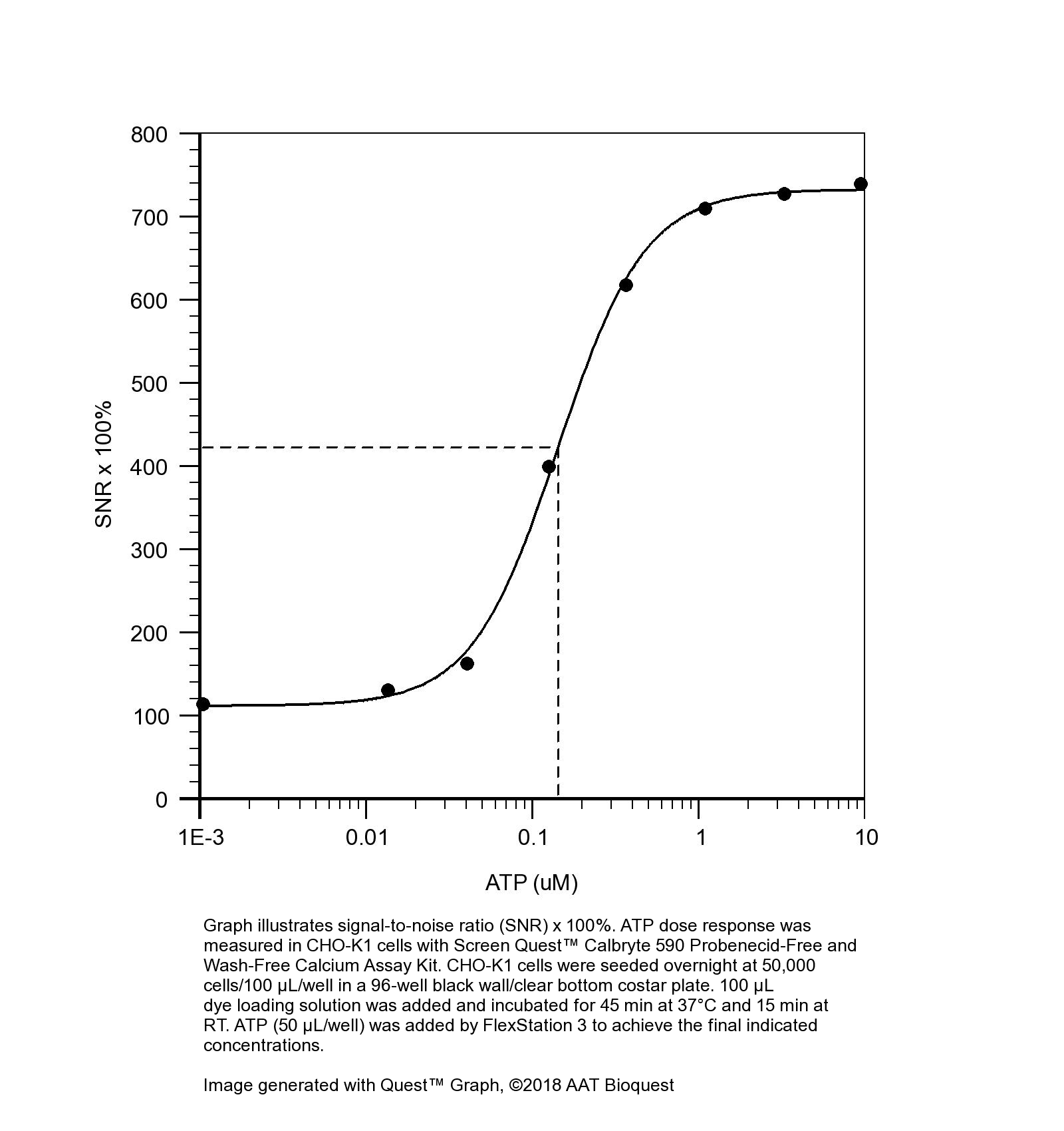
 图1.图表说明了信噪比(SNR)x 100%。 使用Screen Quest Calbryte 590无丙磺舒和无洗涤钙测定试剂盒测量CHO-K1细胞中的ATP剂量反应。 将CHO-K1细胞以50,000个细胞/ 100 µL /孔在96孔黑色板上接种过夜。 加入100 µL的染料加载溶液,在37°C下孵育45分钟,在室温下孵育15分钟。 FlexStation 3添加了ATP(50 µL /孔)以达到最终指示的浓度。 |
相关产品
Calreticulin regulates TGF-β1-induced epithelial mesenchymal transition through modulating Smad signaling and calcium signaling
Authors: Wu, Yanjiao and Xu, Xiaoli and Ma, Lunkun and Yi, Qian and Sun, Weichao and Tang, Liling
Journal: The International Journal of Biochemistry & Cell Biology (2017)
Dexmedetomidine reduces hypoxia/reoxygenation injury by regulating mitochondrial fission in rat hippocampal neurons
Authors: Liu, Jia and Du, Qing and Zhu, He and Li, Yu and Liu, Maodong and Yu, Shoushui and Wang, Shilei
Journal: Int J Clin Exp Med (2017): 6861–6868
Monosialoganglioside 1 may alleviate neurotoxicity induced by propofol combined with remifentanil in neural stem cells
Authors: Lu, Jiang and Yao, Xue-qin and Luo, Xin and Wang, Yu and Chung, Sookja Kim and Tang, He-xin and Cheung, Chi Wai and Wang, Xian-yu and Meng, Chen and Li, Qing and others
Journal: Neural Regeneration Research (2017): 945
Obtaining spontaneously beating cardiomyocyte-like cells from adipose-derived stromal vascular fractions cultured on enzyme-crosslinked gelatin hydrogels
Authors: Yang, Gang and Xiao, Zhenghua and Ren, Xiaomei and Long, Haiyan and Ma, Kunlong and Qian, Hong and Guo, Yingqiang
Journal: Scientific Reports (2017): 41781
Di (2-ethylhexyl) phthalate-induced apoptosis in rat INS-1 cells is dependent on activation of endoplasmic reticulum stress and suppression of antioxidant protection
Authors: Sun, Xia and Lin, Yi and Huang, Qiansheng and Shi, Junpeng and Qiu, Ling and Kang, Mei and Chen, Yajie and Fang, Chao and Ye, Ting and Dong, Sijun
Journal: Journal of cellular and molecular medicine (2015): 581–594
The effect of mitochondrial calcium uniporter on mitochondrial fission in hippocampus cells ischemia/reperfusion injury
Authors: Zhao, Lantao and Li, Shuhong and Wang, Shilei and Yu, Ning and Liu, Jia
Journal: Biochemical and biophysical research communications (2015): 537–542
Fungus induces the release of IL-8 in human corneal epithelial cells, via Dectin-1-mediated protein kinase C pathways.
Authors: Peng, Xu-Dong and Zhao, Gui-Qiu and Lin, Jing and Jiang, Nan and Xu, Qiang and Zhu, Cheng-Cheng and Qu, Jain-Qiu and Cong, Lin and Li, Hui
Journal: International journal of ophthalmology (2014): 441–447
Propofol and remifentanil at moderate and high concentrations affect proliferation and differentiation of neural stem/progenitor cells
Authors: Li, Qing and Lu, Jiang and Wang, Xianyu and others
Journal: Neural regeneration research (2014): 2002
Role of mitochondrial calcium uniporter in regulating mitochondrial fission in the cerebral cortexes of living rats
Authors: Liang, Nan and Wang, Peng and Wang, Shilei and Li, Shuhong and Li, Yu and Wang, Jinying and Wang, Min
Journal: Journal of Neural Transmission (2014): 593–600
Increased expression of cell adhesion molecule 1 by mast cells as a cause of enhanced nerve–mast cell interaction in a hapten-induced mouse model of atopic dermatitis
Authors: Hagiyama, M and Inoue, T and Furuno, T and Iino, T and Itami, S and Nakanishi, M and Asada, H and Hosokawa, Y and Ito, A
Journal: British Journal of Dermatology (2013): 771–778