上海金畔生物科技有限公司代理AAT Bioquest荧光染料全线产品,欢迎访问AAT Bioquest荧光染料官网了解更多信息。
荧光底物Xite Green β-D-半乳糖吡喃糖苷*绿色荧光*
 |
货号 | 14030 | 存储条件 | 在零下15度以下保存, 避免光照 |
| 规格 | 1 mg | 价格 | 2472 | |
| Ex (nm) | Em (nm) | |||
| 分子量 | 494.50 | 溶剂 | DMSO | |
| 产品详细介绍 | ||||
简要概述
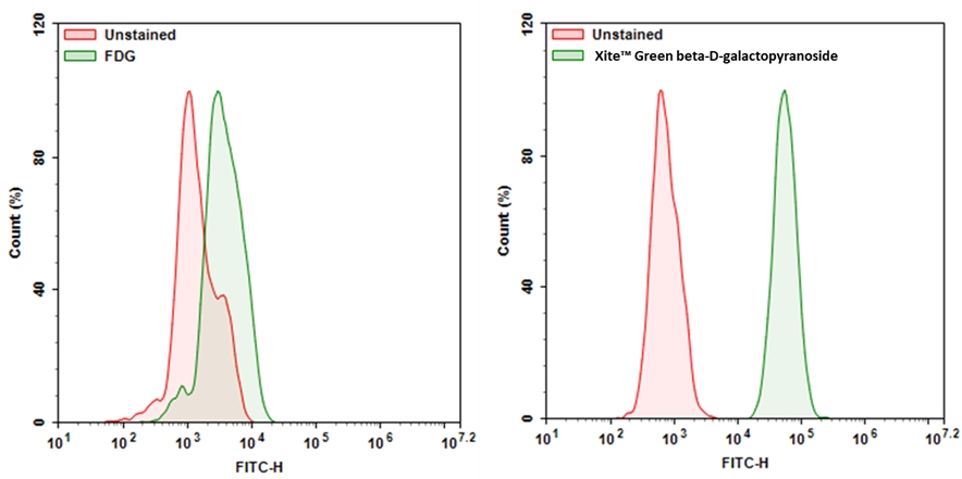
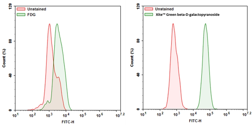
Xite Green β-D-半乳糖吡喃糖苷是β-半乳糖苷酶(β-gal)的荧光底物。与现有的β-半乳糖苷酶底物(例如,常用的FDG)相比,它具有更好的细胞通透性。Xite Green β-D-吡喃半乳糖苷很容易进入细胞,并被β-gal裂解,从而产生强荧光产品Xite Green。强烈荧光的Xite Green可以很好地保留在细胞中,从而易于通过流式细胞仪和荧光显微镜进行检测。Xite Greenβ-D-半乳糖吡喃糖苷提供了一种简单而灵敏的工具来检测β-半乳糖苷酶的活性。Xite Greenβ-D-吡喃半乳糖苷可以作为检测细胞中细胞衰老的工具,因为β-gal已被确定为细胞衰老的可靠标记。金畔生物是AAT Bioquest的中国代理商,为您提供最优质的Xite Green β-D-半乳糖吡喃糖苷。
适用仪器
| 流式细胞仪 | |
| 激发: | 488nm激光 |
| 发射: | 530/30 nm滤波片 |
| 通道: | FITC滤波片组 |
| 荧光显微镜 | |
| 激发: | FITC滤波片 |
| 发射: | FITC滤波片 |
| 推荐孔板: | 黑色透明 |
产品说明书
样品实验方案
以下是我们推荐的方案,仅提供指导。具体实验应根据您的特定需求进行修改。
简要概述
- 根据需要处理样品
- 准备Xite Green β-D-吡喃半乳糖苷工作溶液并将其添加到样品中
- 在37°C下孵育样品15至45分钟
- 使用带有530/30 nm滤光片的流式细胞仪(FITC通道)或带有FITC滤光片组的荧光显微镜检测荧光强度
溶液配制
储备溶液配制
操作步骤
- 根据需要处理样品。
- 处理并用自备的缓冲液(例如DPBS)洗涤细胞。
- 加入Xite Green β-D-吡喃半乳糖苷工作溶液15-45分钟,然后在37°C的培养箱中培养样品。
注意:孵育的最佳时间需要通过实验确定。 - 取出工作溶液并用自备的缓冲液洗涤细胞。
- 将细胞重悬在自备的缓冲液中,并使用流式细胞仪使用530/30 nm滤光片(FITC通道)或带有FITC滤光片组的荧光显微镜检测荧光强度。
图示
 图1.用Xite Green β-D-吡喃半乳糖苷测量β-gal的表达。将9L-LacZ细胞(过度表达β-gal的细胞)与Xite Green β-D-半乳糖吡喃糖苷或FDG在37°C孵育30分钟。使用NovoCyte流式细胞仪(ACEA Biosciences)通过FITC通道获取信号。 |
参考文献
Cellular and cytoskeletal alterations of scleral fibroblasts in response to glucocorticoid steroids.
Authors: Bogarin, Thania and Saraswathy, Sindhu and Akiyama, Goichi and Xie, Xiaobin and Weinreb, Robert N and Zheng, Jie and Huang, Alex S
Journal: Experimental eye research (2019): 107774
Novel fluorescent probe for rapid and ratiometric detection of β-galactosidase and live cell imaging.
Authors: Chen, Xiangzhu and Zhang, Xueyan and Ma, Xiaodong and Zhang, Yuanyuan and Gao, Gui and Liu, Jingjing and Hou, Shicong
Journal: Talanta (2019): 308-313
SA-β-Galactosidase-Based Screening Assay for the Identification of Senotherapeutic Drugs.
Authors: Fuhrmann-Stroissnigg, Heike and Santiago, Fernando E and Grassi, Diego and Ling, YuanYuan and Niedernhofer, Laura J and Robbins, Paul D
Journal: Journal of visualized experiments : JoVE (2019)
Targeting senescence improves angiogenic potential of adipose-derived mesenchymal stem cells in patients with preeclampsia.
Authors: Suvakov, Sonja and Cubro, Hajrunisa and White, Wendy M and Butler Tobah, Yvonne S and Weissgerber, Tracey L and Jordan, Kyra L and Zhu, Xiang Y and Woollard, John R and Chebib, Fouad T and Milic, Natasa M and Grande, Joseph P and Xu, Ming and Tchkonia, Tamara and Kirkland, James L and Lerman, Lilach O and Garovic, Vesna D
Journal: Biology of sex differences (2019): 49
Tumor cell escape from therapy-induced senescence.
Authors: Saleh, Tareq and Tyutyunyk-Massey, Liliya and Murray, Graeme F and Alotaibi, Moureq R and Kawale, Ajinkya S and Elsayed, Zeinab and Henderson, Scott C and Yakovlev, Vasily and Elmore, Lynne W and Toor, Amir and Harada, Hisashi and Reed, Jason and Landry, Joseph W and Gewirtz, David A
Journal: Biochemical pharmacology (2019): 202-212
Fluorescent probes for selective protein labeling in lysosomes: a case of α-galactosidase A.
Authors: Bohl, Cornelius and Pomorski, Adam and Seemann, Susanne and Knospe, Anne-Marie and Zheng, Chaonan and Krężel, Artur and Rolfs, Arndt and Lukas, Jan
Journal: FASEB journal : official publication of the Federation of American Societies for Experimental Biology (2017): 5258-5267
Identification of a β-galactosidase transgene that provides a live-cell marker of transcriptional activity in growing oocytes and embryos.
Authors: Edwards, Nicole and Farookhi, Riaz and Clarke, Hugh J
Journal: Molecular human reproduction (2015): 583-93
Characterization of functional capacity of adult ventricular myocytes in long-term culture.
Authors: Liu, Shi J
Journal: International journal of cardiology (2013): 1923-36
Abrupt and dynamic changes in gene expression revealed by live cell arrays.
Authors: Walling, Maureen A and Shi, Hua and Shepard, Jason R E
Journal: Analytical chemistry (2012): 2737-44
Live-cell imaging visualizes frequent mitotic skipping during senescence-like growth arrest in mammary carcinoma cells exposed to ionizing radiation.
Authors: Suzuki, Masatoshi and Yamauchi, Motohiro and Oka, Yasuyoshi and Suzuki, Keiji and Yamashita, Shunichi
Journal: International journal of radiation oncology, biology, physics (2012): e241-50
